Eaton-Peabody Laboratories
3D Virtual Model of the Round Window Area in Humans
About the Round Window Model
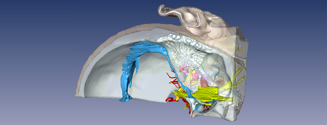
The Eaton-Peabody Laboratories have developed a 3D virtual model of the anatomy of the round window membrane and adjacent cochlear structures in the human. Knowledge of the spatial relationship of these structures has implications for cochlear implantation, wherein preservation of residual hearing is an important aim in some cases.
Archival, 20 micron, celloidin embedded sections from a 14-year-old male were used. Each and every section through the round window was stained, digitized, and imported into a 3D software program (Amira, version 3.1). A total of 201 number of sections were used to generate the model.
The resulting 3D model is a surface rendering of structures of interest, including the round window, basilar membrane, osseous spiral lamina, scala media, spiral ligament, cochlear aqueduct, inferior cochlear vein and related venous structures, scala vestibuli, scala tympani, and ductus reuniens.
Installation by Operating System
- Download and uncompress the setup package
- Double click on the setup.exe file and follow the instructions on the wizard to install
- The program will automatically run after the installation
- Please refer to the user guide for how to use this software
- Download and uncompress the setup package
- Double click on the setup.exe file and follow the instructions on the wizard to install
- The program will automatically run after the installation
This model was developed by Haobing Wang, Clarinda Northrop, Barbara Burgess, Saumil Merchant, MD, and Joseph B. Nadol, Jr., MD, of Mass Eye and Ear.


