Imaging Services
X-Ray and Fluoroscopy
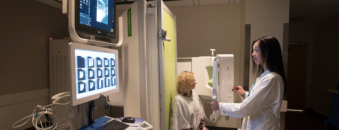
X-ray, also known as radiography, is the oldest form of diagnostic imaging. X-ray imaging is a fast and efficient diagnostic method that aids physicians in viewing internal bodily structures, thereby enabling them to diagnose both disease and injury.
X-ray technology involves passing an X-ray beam through a part of the body to ultimately produce images of internal structures. As X-rays penetrate the body, they are absorbed in varying amounts by different tissues.
Fluoroscopy is a technique used to capture X-ray images in motion. During a fluoroscopic procedure, a radiologist transmits a low energy X-ray beam (lower energy than traditional X-rays) through the patient onto a plate or carriage. The plate is connected to an image intensifier that is in turn connected to a video screen. The resulting image projected on the video screen is a live X-ray movie of internal organs in motion in a specific region of the body.
Information for Patients
Fluoroscopy is used during these examinations: barium swallow, modified barium swallow, dacrocystogram, sialogram, velopharyngeal insufficiency (VPI) and nasogastric tube placement.
We use techniques during our exams to significantly reduce the dose of radiation.
If you are seeing one of the physicians at Massachusetts Eye and Ear your appointment will be scheduled through the office. Otherwise you can call 617-573-3555 to schedule any of your imaging studies through the Radiology department. Our staff have many years of experience in helping you prepare for your Radiology exam procedure. We will call you prior to your appointment, if we need further information of the imaging study or procedure.


