
December 2, 2020Press Release
Harvard Medical School Scientists Reverse Age-Related Vision Loss, Eye Damage from Glaucoma in Mice
Media Contacts
Ryan Jaslow | Mass Eye and Ear
617-573-4385 | Ryan_Jaslow@meei.harvard.edu
Ekaterina Pesheva | Harvard Medical School
617-432-0441 | Ekaterina_Pesheva@hms.harvard.edu
At-a-glance:
- Proof-of-concept study represents first successful attempt to reverse the aging clock in animals through epigenetic reprogramming.
- Scientists turned on embryonic genes to reprogram cells of mouse retinas.
- Approach reversed glaucoma-induced eye damage in animals.
- Approach also restored age-related vision loss in elderly mice.
- Work spells promise for using same approach in other tissues, organs beyond the eyes.
- Success sets stage for treatment of various age-related diseases in humans.
Harvard Medical School scientists have successfully restored vision in mice by turning back the clock on aged eye cells in the retina to recapture youthful gene function.
The team’s work, described Dec. 2 in Nature, represents the first demonstration that it may be possible to safely reprogram complex tissues, such as the nerve cells of the eye, to an earlier age.
In addition to resetting the cells’ aging clock, the researchers successfully reversed vision loss in animals with a condition mimicking human glaucoma, a leading cause of blindness around the world.
The achievement represents the first successful attempt to reverse glaucoma-induced vision loss, rather than merely stem its progression, the team said.
If replicated through further studies, the approach could pave the way for therapies to promote tissue repair across various organs and reverse aging and age-related diseases in humans.
“Our study demonstrates that it’s possible to safely reverse the age of complex tissues such as the retina and restore its youthful biological function,” said senior author David Sinclair, PhD, professor of genetics in the Blavatnik Institute at Harvard Medical School, co-director of the Paul F. Glenn Center for Biology of Aging Research at HMS and an expert on aging.
Sinclair and colleagues caution that the findings remain to be replicated in further studies, including in different animal models, before any human experiments. Nonetheless, they add, the results offer a proof of concept and a pathway to designing treatments for a range of age-related human disease.
“If affirmed through further studies, these findings could be transformative for the care of age-related vision diseases like glaucoma and to the fields of biology and medical therapeutics for disease at large,” Sinclair said.
For their work, the team used an adeno-associated virus (AAV) as a vehicle to deliver into the retinas of mice three youth-restoring genes—Oct4, Sox2 and Klf4—that are normally switched on during embryonic development. The three genes, together with a fourth one, which was not used in this work, are collectively known as Yamanaka factors.
The treatment had multiple beneficial effects on the eye. First, it promoted nerve regeneration following optic-nerve injury in mice with damaged optic nerves. Second, it reversed vision loss in animals with a condition mimicking human glaucoma. And third, it reversed vision loss in aging animals without glaucoma.
The team’s approach is based on a new theory about why we age. Most cells in the body contain the same DNA molecules but have widely diverse functions. To achieve this degree of specialization, these cells must read only genes specific to their type. This regulatory function is the purview of the epigenome, a system of turning genes on and off in specific patterns without altering the basic underlying DNA sequence of the gene.
This theory postulates that changes to the epigenome over time cause cells to read the wrong genes and malfunction––giving rise to diseases of aging. One of the most important changes to the epigenome is DNA methylation, a process by which methyl groups are tacked onto DNA. Patterns of DNA methylation are laid down during embryonic development to produce the various cell types. Over time, youthful patterns of DNA methylation are lost, and genes inside cells that should be switched on get turned off and vice versa, resulting in impaired cellular function. Some of these DNA methylation changes are predictable and have been used to determine the biologic age of a cell or tissue.
Yet, whether DNA methylation drives age-related changes inside cells has remained unclear. In the current study, the researchers hypothesized that if DNA methylation does, indeed, control aging, then erasing some of its footprints might reverse the age of cells inside living organisms and restore them to their earlier, more youthful state.
Past work had achieved this feat in cells grown in laboratory dishes but fell short of demonstrating the effect in living organisms.
The new findings demonstrate that the approach could be used in animals as well.
Overcoming an important hurdle
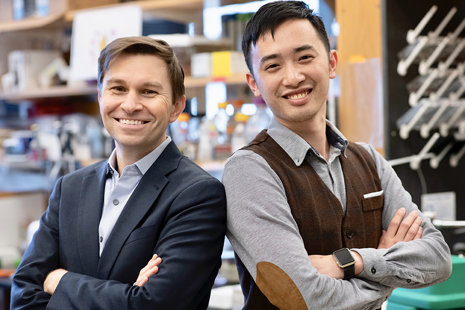
Lead study author, Yuancheng Lu, PhD, research fellow in genetics at HMS and a former doctoral student in Sinclair’s lab, developed a gene therapy that could safely reverse the age of cells in a living animal.
Lu’s work builds on the Nobel Prize winning discovery of Shinya Yamanaka, who identified the four transcription factors, Oct4, Sox2, Klf4, c-Myc, that could erase epigenetics markers on cells and return these cells to their primitive embryonic state from which they can develop into any other type of cell.
Subsequent studies, however, showed two important setbacks. First, when used in adult mice, the four Yamanaka factors could also induce tumor growth, rendering the approach unsafe. Second, the factors could reset the cellular state to the most primitive cell state, thus completely erasing a cell’s identity.
Lu and colleagues circumvented these hurdles by slightly modifying the approach. They dropped the gene c-Myc and delivered only the remaining three Yamanaka genes, Oct4, Sox2 and Klf4. The modified approach successfully reversed cellular aging without fueling tumor growth or losing their identity.
Gene therapy applied to optic nerve regeneration
In the current study, the researchers targeted cells in the central nervous system because it is the first part of body affected by aging. After birth, the ability of the central nervous system to regenerate declines rapidly.
To test whether the regenerative capacity of young animals could be imparted to adult mice, the researchers delivered the modified three-gene combination via an AAV into retinal ganglion cells of adult mice with optic nerve injury.
For the work, Lu and Sinclair partnered with Zhigang He, PhD, HMS professor of neurology and of ophthalmology at Boston Children’s Hospital, who studies optic nerve and spinal cord neuro-regeneration.
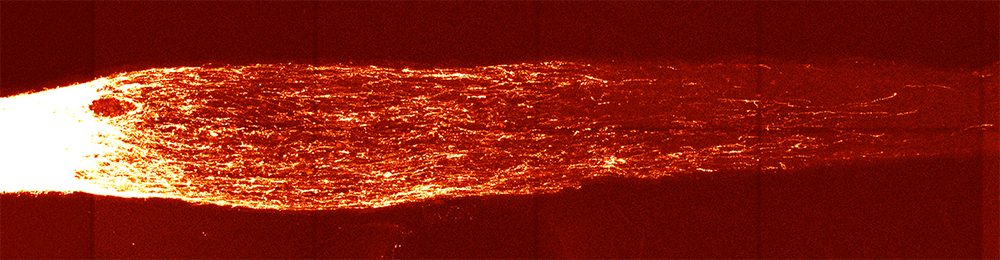
The treatment resulted in a two-fold increase in the number of surviving retinal ganglion cells after the injury and a five-fold increase in nerve regrowth.
“At the beginning of this project, many of our colleagues said our approach would fail or would be too dangerous to ever be used,” said Lu. “Our results suggest this method is safe and could potentially revolutionize the treatment of the eye and many other organs affected by aging.”
Reversal of glaucoma and age-related vision loss
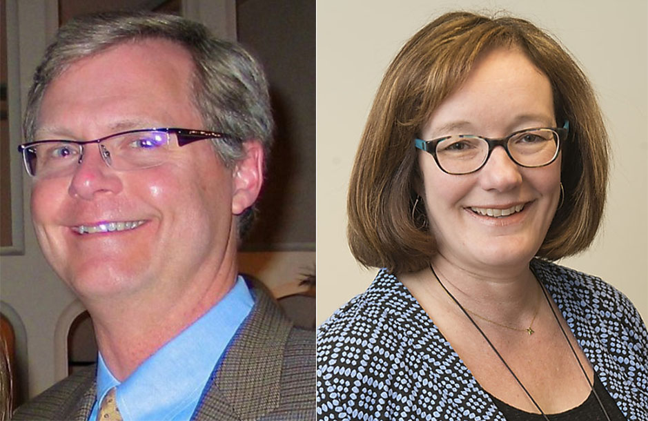
Following the encouraging findings in mice with optic nerve injuries, the team partnered with colleagues at Schepens Eye Research Institute of Massachusetts Eye and Ear Bruce Ksander, PhD, HMS associate professor of ophthalmology, and Meredith Gregory-Ksander, PhD, HMS assistant professor of ophthalmology. They planned two sets of experiments: one to test whether the three-gene cocktail could restore vision loss due to glaucoma and another to see whether the approach could reverse vision loss stemming from normal aging.
In a mouse model of glaucoma, the treatment led to increased nerve cell electrical activity and a notable increase in visual acuity, as measured by the animals’ ability to see moving vertical lines on a screen. Remarkably, it did so after the glaucoma-induced vision loss had already occurred.
“Regaining visual function after the injury occurred has rarely been demonstrated by scientists,” Ksander said. “This new approach, which successfully reverses multiple causes of vision loss in mice without the need for a retinal transplant, represents a new treatment modality in regenerative medicine.”
The treatment worked similarly well in elderly, 12-month-old mice with diminishing vision due to normal aging. Following treatment of the elderly mice, the gene expression patterns and electrical signals of the optic nerve cells were similar to young mice, and vision was restored.
When the researchers analyzed molecular changes in treated cells, they found reversed patterns of DNA methylation—an observation suggesting that DNA methylation is not a mere marker or a bystander in the aging process, but rather an active agent driving it.
“What this tells us is the clock doesn’t just represent time—it is time,” said Sinclair. “If you wind the hands of the clock back, time also goes backward.”
“Currently, treatment strategies for many age-related eye diseases—like glaucoma—are focused on slowing disease progression and preserving the patient’s remaining vision. This research is very promising, as it may ultimately lead to a new class of therapeutics that could restore vision that has already been lost,” said Joan W. Miller, MD, Chief of Ophthalmology at Mass Eye and Ear, Massachusetts General Hospital, and Ophthalmologist-in Chief at Brigham and Women’s Hospital, and Chair of Ophthalmology and David Glendenning Cogan Professor of Ophthalmology at Harvard Medical School.
The researchers said that if their findings are confirmed in further animal work, they could initiate clinical trials within two years to test the efficacy of the approach in people with glaucoma. Thus far, the findings are encouraging, researchers said. In the current study, a one-year, whole-body treatment of mice with the three-gene approach showed no negative side effects.
Other authors on the paper include Benedikt Brommer, Xiao Tian, Anitha Krishnan, Margarita Meer, Chen Wang, Daniel Vera, Qiurui Zeng, Doudou Yu, Michael Bonkowski, Jae-Hyun Yang, Songlin Zhou, Emma Hoffmann, Margarete Karg, Michael Schultz, Alice Kane, Noah Davidsohn, Ekaterina Korobkina, Karolina Chwalek, Luis Rajman, George Church, Konrad Hochedlinger, Vadim Gladyshev, Steve Horvath and Morgan Levine.
This work was supported in part by a Harvard Medical School Epigenetics Seed Grant and Development Grant, The Glenn Foundation for Medical Research, Edward Schulak, the National Institutes of Health (grants R01AG019719, R37AG028730, R01EY026939, R01EY021526, R01AG067782, R01GM065204, R01AG065403, R01EY025794, R24EY028767 and R21EY030276), and the St. Vincent de Paul Foundation.
Relevant disclosures: David Sinclair is a consultant to, inventor of patents licensed to, board member and equity owner of Iduna Therapeutics, a Life Biosciences company developing epigenetic reprogramming therapies, and an unpaid consultant to Zymo Research, an epigenetic tools company. Yuancheng Lu, Luis Rajman and Steve Horvath are equity owners of Iduna Therapeutics. George Church and Noah Davidsohn are co-founders of Rejuvenate Bio. Additional Sinclair and Church disclosures are at http://arep.med.harvard.edu/gmc/tech.html and https://genetics.med.harvard.edu/sinclair/people/sinclair-other.php.
About Harvard Medical School
Harvard Medical School has more than 11,000 faculty working in the 11 basic and social science departments comprising the Blavatnik Institute and at the 15 Harvard-affiliated teaching hospitals and research institutes: Beth Israel Deaconess Medical Center, Boston Children’s Hospital, Brigham and Women’s Hospital, Cambridge Health Alliance, Dana-Farber Cancer Institute, Harvard Pilgrim Health Care Institute, Hebrew SeniorLife, Joslin Diabetes Center, Judge Baker Children’s Center, Massachusetts Eye and Ear/Schepens Eye Research Institute, Massachusetts General Hospital, McLean Hospital, Mount Auburn Hospital, Spaulding Rehabilitation Network and VA Boston Healthcare System.
About Mass Eye and Ear
Massachusetts Eye and Ear, founded in 1824, is an international center for treatment and research and a teaching hospital of Harvard Medical School. A member of Mass General Brigham, Mass Eye and Ear specializes in ophthalmology (eye care) and otolaryngology–head and neck surgery (ear, nose and throat care). Mass Eye and Ear clinicians provide care ranging from the routine to the very complex. Also home to the world's largest community of hearing and vision researchers, Mass Eye and Ear scientists are driven by a mission to discover the basic biology underlying conditions affecting the eyes, ears, nose, throat, head and neck and to develop new treatments and cures. In the 2020–2021 “Best Hospitals Survey,” U.S. News & World Report ranked Mass Eye and Ear #4 in the nation for eye care and #6 for ear, nose and throat care. For more information about life-changing care and research at Mass Eye and Ear, visit our blog, Focus, and follow us on Instagram, Twitter and Facebook.
About Harvard Medical School Department of Ophthalmology
The Harvard Medical School Department of Ophthalmology is one of the leading and largest academic departments of ophthalmology in the nation. Composed of nine affiliates (Massachusetts Eye and Ear, which is home to Schepens Eye Research Institute; Massachusetts General Hospital; Brigham and Women’s Hospital; Boston Children’s Hospital; Beth Israel Deaconess Medical Center; Joslin Diabetes Center/Beetham Eye Institute; Veterans Affairs Boston Healthcare System; Veterans Affairs Maine Healthcare System; and Cambridge Health Alliance) and several international partners, the department draws upon the resources of a global team to pursue a singular goal—eradicate blinding diseases so that all children born today will see throughout their lifetimes. Formally established in 1871, the department is committed to its three-fold mission of providing premier clinical care, conducting transformational research, and providing world-class training for tomorrow’s leaders in ophthalmology.