
Eaton-Peabody Laboratories
Histology Core
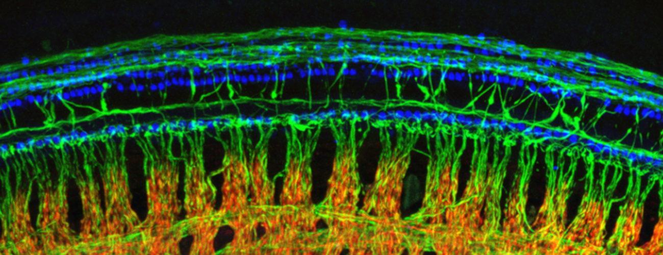
The Histology Core of the Eaton-Peabody Laboratories (EPL) at Mass. Eye and Ear strives to develop and refine techniques for histological analysis, particularly as applied to the study of the inner ear. Below you will find several resources made publicly available by the EPL team.
Cochlear Dissection for Whole Mount Immunostaining
We have prepared a 35-minute cochlear dissection video illustrating how to harvest the entire organ of Corti from an adult mouse ear and prepare it for viewing with high-power objectives in order to produce confocal images. If you find this video useful, please email comments to M. Charles Liberman, PhD.
View Dissection Summary (PDF)View PDF Of Cochlear Whole Mount Dissection Instructions
Mapping Cochlear Length to Cochlear Frequency
Once the organ of Corti has been dissected into component pieces, reconstructing the total length and converting cochlear location to cochlear frequency is simplified with this ImageJ plug-in. It fits spline curves to user-defined arcs superimposed on the cochlear pieces and displays the locations of half-octave intervals of cochlear frequency. Mapping functions for a variety of animals, including humans, are built-in options. It can also automatically calculate the frequencies at different points along the cochlea. The plug-in can also calculate the frequency at additional points of interest, or divide the total length into equal segments for haircell counting.
Please read the User Manual for step by step instructions on how to install and use the plug-in.
Plastic Embedding and Thick Sectioning for Temporal Bone Histopathology
When high-resolution analysis of all structures of the inner ear is required, plastic embedding of aldehyde fixed and osmium post-fixed tissue provides much better morphological preservation than conventional paraffin embedding. Material can be sectioned at 40 um for light-microscopic study, and then re-embedded for ultrathin sectioning and electron microscopic examination. Fixed tissue is osmicated, decalcified, dehydrated, and infiltrated with plastic via a graded series of propylene oxide : araldite solutions, before being hardened in a 60 degree C oven. Cured blocks may be sectioned at thicknesses from ultrathin to 100 um.